Specifically designed for Image Scanning Microscopy, compatible with all scanning techniques.
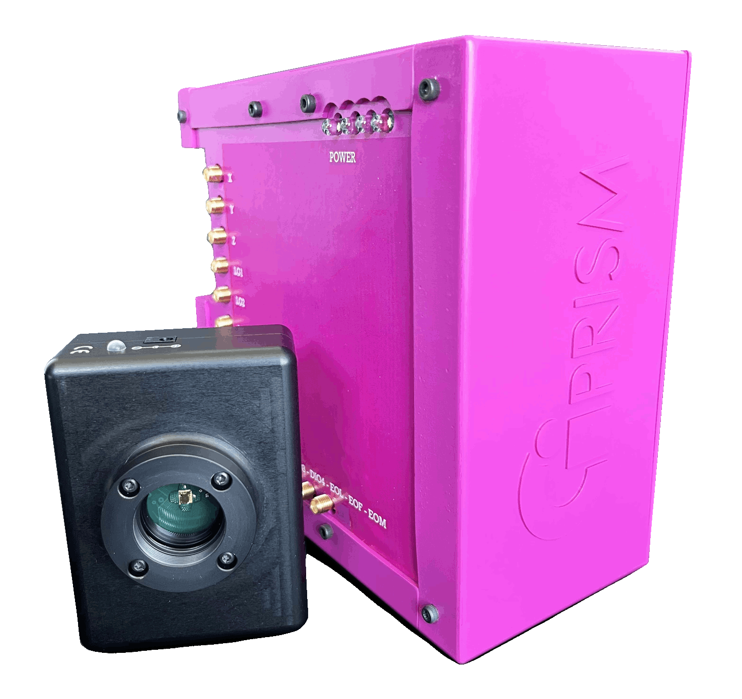
CARMA has been specifically designed by our team for PRISM technology. It is conceived for Image Scanning Microscopy and it is also compatible with most laser scanning setups.
Originally used in
DiasproLab and
VicidominiLab at the
Istituto Italiano di Tecnologia,
CARMA wants to offer the best imaging experience for ISM imaging. It enables for easy and plug-and-play multi-sensor imaging via simultaneous independent acquisition channels and it integrates the best code to precisely control multiple digital and analog devices.
Parallel asynchronous acquisition from SPAD arrays, APDs and PMTs.
Real-time FPGA-based system for the best image processing.
Multi-channel Analog/Digital control for maximum versatility.
Easy control of Galvo-mirrors, Piezo Stages, Shutters, Lasers, AODs, AOMs.
A field-programmable gate array (FPGA) is an integrated circuit that can be used to implement any logical function that an application-specific integrated circuit (ASIC) could perform.
Among the many advantages of using an FPGA-based control system, we can highlight the possibility to have fast and efficient hardware control, massive parallel data processing, real-time applications, tailored software/hardware configuration, updating and remote configuring capabilities.
Our FPGA control unit offers complete control over the synchronization and timing of all input/output signals and operations and the possibility to perform custom onboard real-time decisions executed with hardware-timed speed and reliability.
The digital lines of the FPGA can be configured as inputs, outputs, counter/timers, PWM, flexible encoder inputs, or bus with specialized communication protocols. Our team is thriving to offer the best tailored solution for ISM as well as for your microscopy setup.
Operating a laser scanning microscope requires precise and reliable control of numerous optomechanical devices, including Galvo-mirrors, motorized positioning stages, laser gates, shutters, acusto-optic tunable filters (AOTF), and acusto-optic modulators (AOM). CARMA offers a complete suite for Image Scanning Microscopy setups and it is also compatible with most common OEMs components.
Galvo-mirrors are used to scan the laser beam(s) across the sample and to de-scan the fluorescence photons that is then collected by the imaging sensor(s). CARMA allows to drive most common XY Galvo Scanners with dedicated analog signal (in the range -10V +10V) or via PWM signals.
Motorized piezo stages are usually used for precise positioning of the sample (or to move objective lenses along the optical axis to select the focal plane). Numerous types of motorized stages can be controlled via dedicated driver by using an analog signal from the CARMA control unit.
Other devices that can be controlled by CARMA include but are not limited to laser gates and shutters (e.g. to allow/block laser lines and to directly control laser intensity), AOTFs, AOMs, and shutters. If needed, tailored analog/digital signals can be provided.
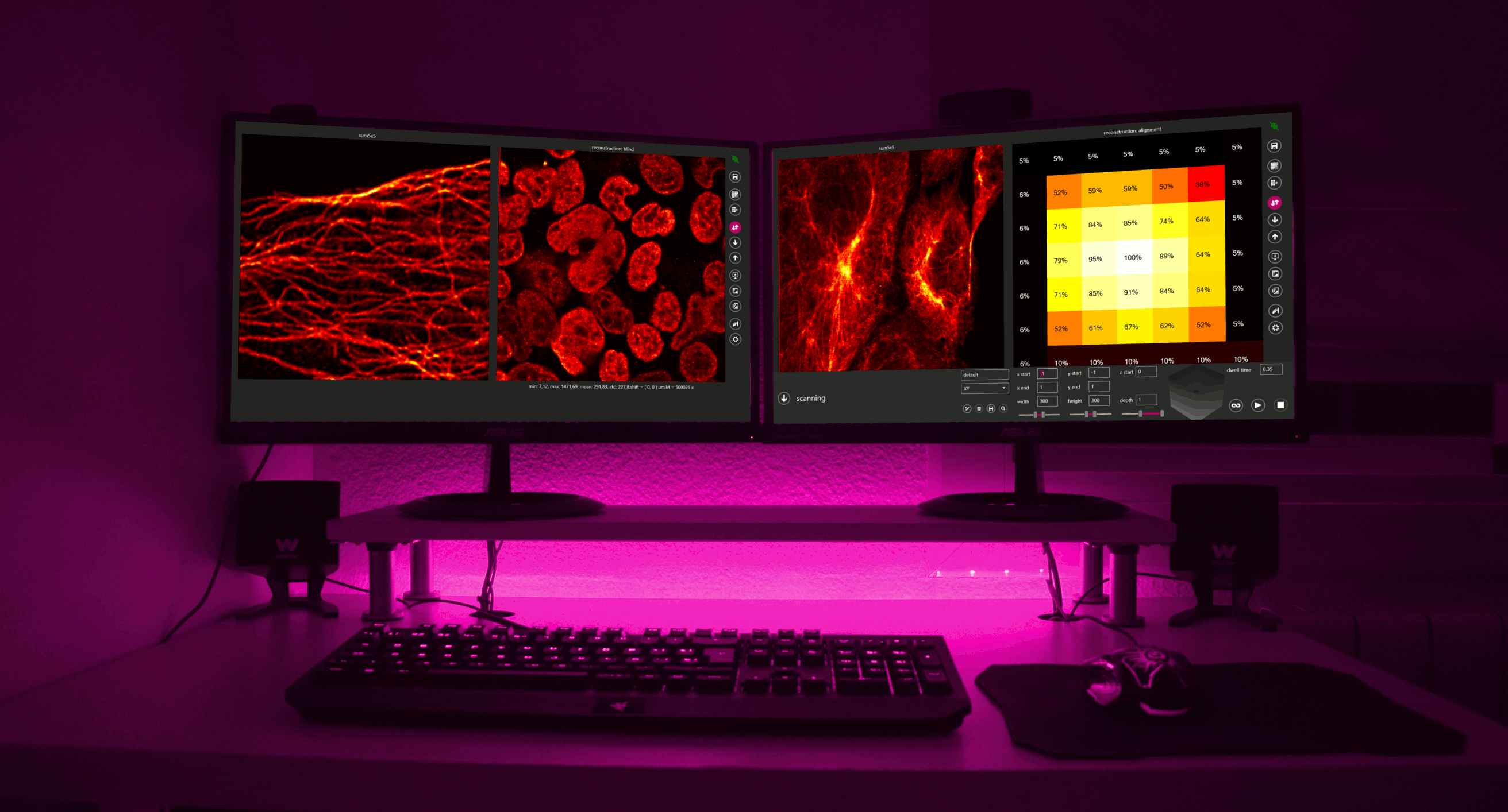
CARMA offers access to the most advanced image acquisition and processing for Image Scanning Microscopy. It enables for parallel and asynchronous
acquisition from up to 50 channels (!) and it is naturally designed to work with SPAD array detectors. Its best performance can be obtained when used with PRISM technology (PRISM-Light, PRISM-Addon, PRISM-Desktop), but it can also manage traditional single elements SPADs, APDs and PMTs, as well as most common scientific imaging cameras.
Single-Photon Avalanche Diodes (SPADs) are popular in optical microscopy thanks to their great reliability, robustness, ease of operation, high detection efficiency, and low timing jitter. They offer individual photons imaging capabilities, with unmatched photon counting and time-resolved performance. Silicon SPADs are typically used to detect photons in the visible wavelength range, from 400 to 1000 nm, while SPADs based on InGaAs/InP are useful for the near-infrared region (from 900 to 1700 nm).
The possibility to easily integrate single SPAD sensors into SPAD arrays has brought to the developments of tailored detectors able to suit the need of most advanced optical microscopy techniques. By replacing the typical single-element sensor used in laser scanning microscopy with a SPAD array, our team revolutionized the world of optical microscopy implementing a natural design of Image Scanning Microscopy capable of extraordinary performance in terms of spatial and temporal resolution.
Photomultiplier tubes (PMTs) represent the current state-of-the-art for confocal microscopy. By exploiting the photoelectric effect, these single-element detectors natively generate a signal that is proportional to the number of photons detected. The best performance in terms of quantum efficiency in the visible range is achieved with Gallium arsenide phosphide cathodes (GaAsP). Despite their high sensitivity, they can't compare with the single photon imaging capabilities offered by SPADs.
Traditionally representing the silicon counterpart of PMTs, Avalanche Photodiodes (APDs) are also very well established in microscopy. They typically offer greater sensitivity for longer wavelengths (500 to 1100 nm) compared to PMTs. While they can be adopted for single-photon detection, they're not naturally designed for time-resolved measurements as SPADs. Recent advances in sensor research and development brought to life the hybrid sensors which aim at combining the sensitivity of PMTs with the photon counting capabilities offered by APDs.
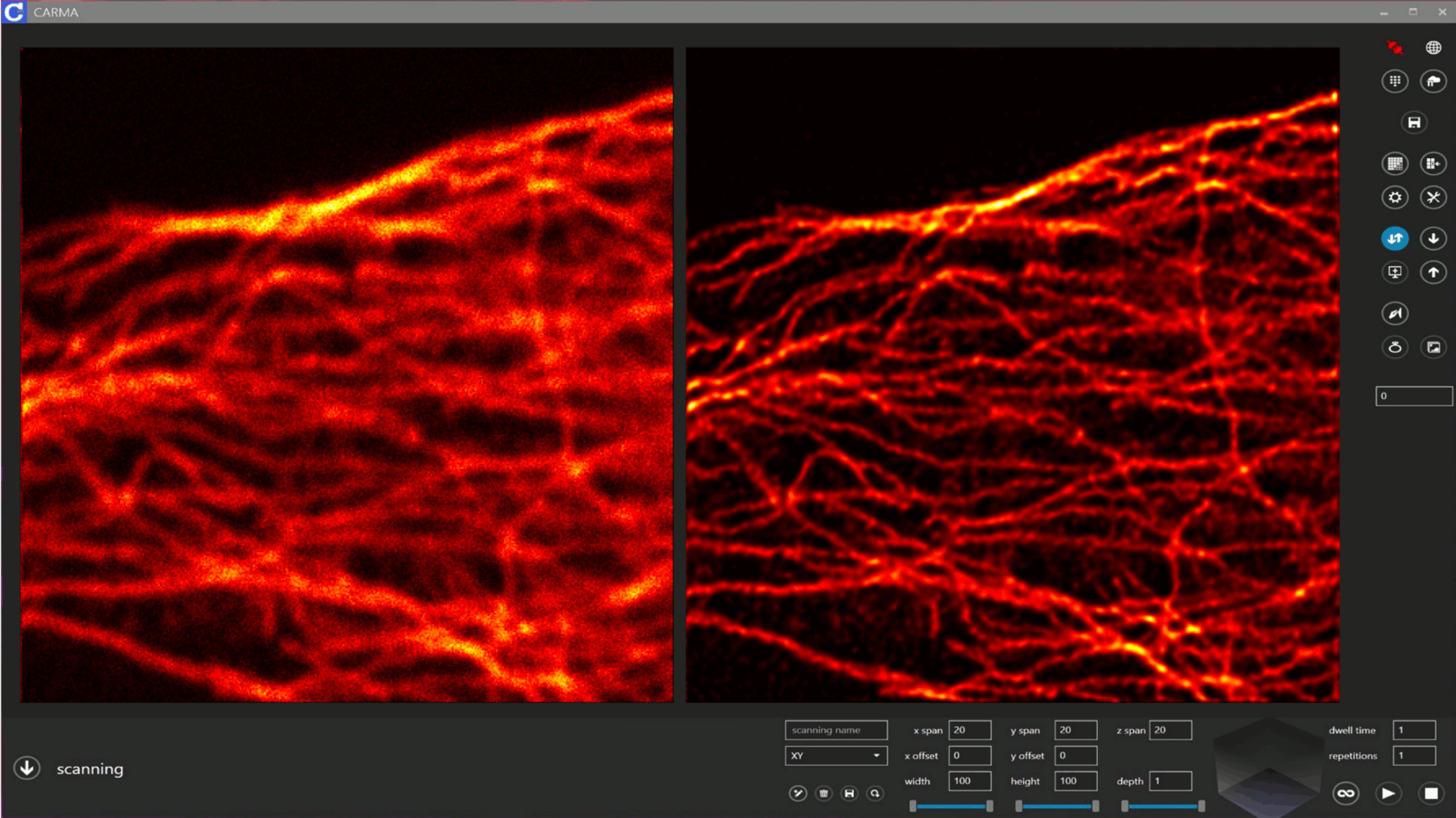
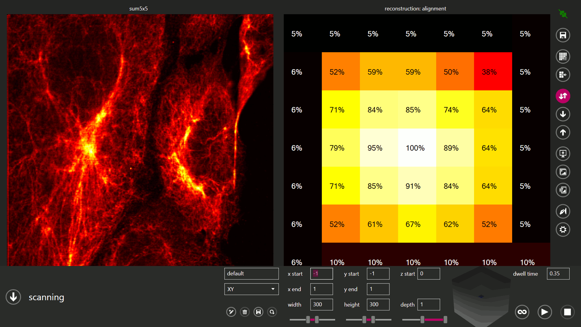
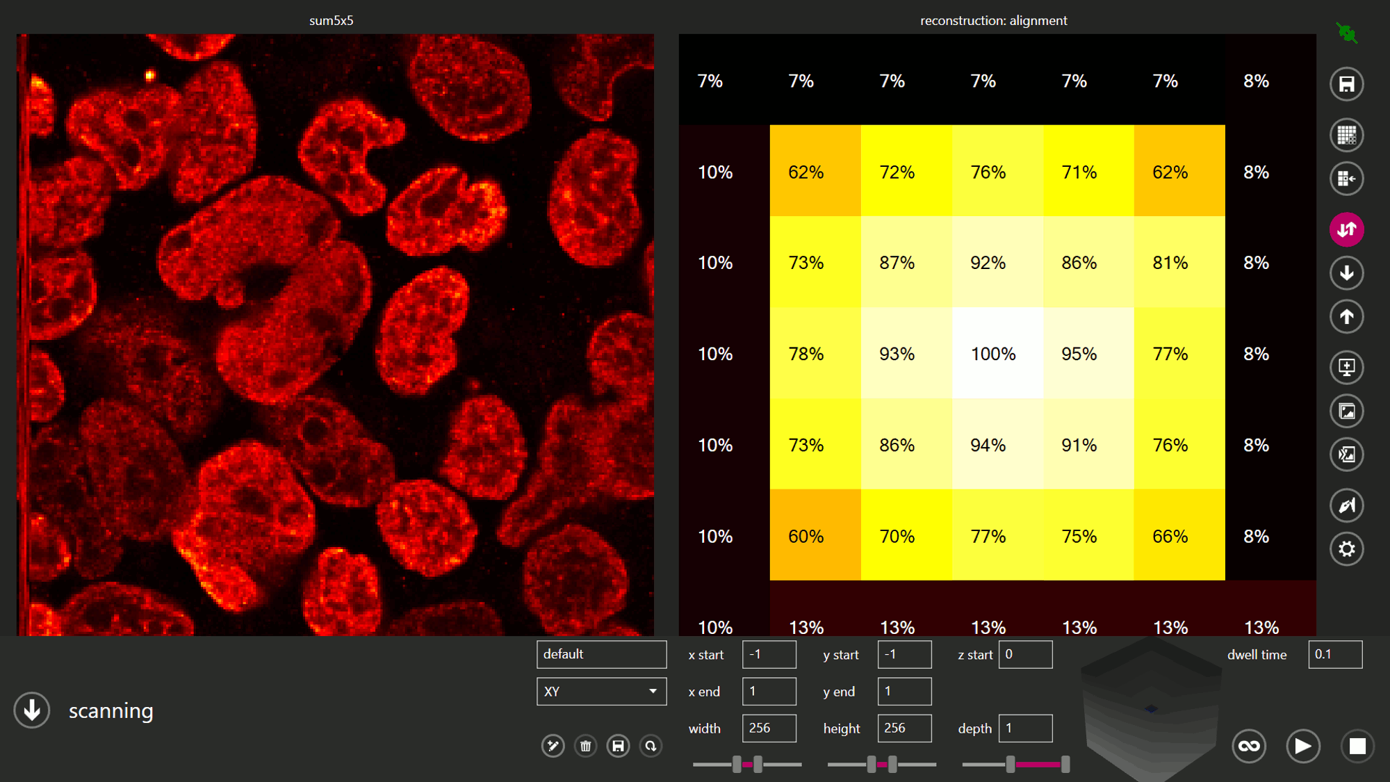
CARMA software can manage all the high-level tasks related to image acquisition, processing and visualization: it offer an intuitive and easy to use user interface for controlling the microscope and setting the parameters for each experiment, it integrates all the image processing and computation algorithms required to create the final image (e.g. Pixel Reassignment), and it implements the best practices for data storage.
Data from each measurement can be saved using most common image file extensions as well as in raw format in order to perform image processing with external data analysis software (i.e. ImageJ). In addition, measurements can be saved directly in Python or MATLAB® files, each containing all the metadata related to the acquired image (pixel dwell-time, pixel size, etc.).
The user interface provides all the standard tools to set the imaging field of view, scanning parameters, detectors configurations, as well as the parameters dedicated to ISM image reconstruction. CARMA also runs a fast computing library specifically designed to exploit parallel computing (GPU processing) enabling for an improved performance when performing live image reconstruction algorithms and deconvolution algorithms.